Looking Beyond Liver Stiffness ...Understanding Fontan-associated liver disease

By Dr James Blackwell, Lecturer in Physics The Fontan procedure, an open-heart surgery, has greatly improved survival for children born with complex congenital heart disease involving only one functional ventricle. However, the altered circulation created by the procedure can also have long-term effects on other organs, particularly the liver.
In a Fontan circulation, systemic venous blood (oxygen-poor blood) is directed towards the lungs without the assistance of a pumping ventricle. This results in chronically elevated venous pressure and reduced cardiac output. Over time, the liver can become congested and develop progressive fibrosis (thickening and scarring of connective tissue), a condition known as Fontan-Associated Liver Disease (FALD) [1,2]. Assessing FALD can be difficult because patients may have relatively few symptoms, routine blood tests do not always reflect the severity of liver disease, and fibrosis can be unevenly distributed throughout the liver.
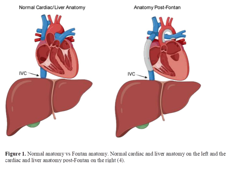
Photo Credit: Gumm AJ, Rand EB. Fontan-Associated Liver Disease. The Journal of Pediatrics. 2025;277. https://doi.org/10.1016/j.jpeds.2024.114389
FALD can progress to cirrhosis and, in advanced cases, may lead to serious complications, making lifelong monitoring important for patients with Fontan circulation. Liver biopsy remains an important reference for assessing fibrosis, but it is invasive and only samples a small area of the liver. This is particularly relevant in FALD, where fibrosis can be unevenly distributed. This has increased interest in non-invasive techniques that can be repeated during long-term follow-up.
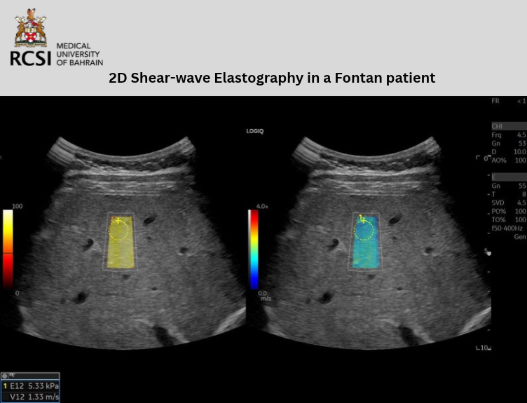
Our collaborative research team has been investigating whether two-dimensional shear-wave elastography (2D-SWE) can help us better understand and monitor these changes. 2D-SWE is an ultrasound technique that generates small mechanical vibrations, known as shear waves, within the tissue. These waves travel faster through stiffer tissue, such as tissue affected by fibrosis or scarring, allowing the liver's stiffness to be estimated non-invasively.
In many chronic liver diseases, increased liver stiffness is associated with fibrosis. Fontan physiology complicates interpretation, as venous congestion can also increase liver stiffness. A high stiffness measurement, therefore, does not necessarily represent fibrosis alone. Our recent narrative review in Abdominal Radiology examined the evidence for 2D-SWE in FALD and highlighted both the influence of hepatic congestion and the lack of validated liver-stiffness thresholds specifically for this population [3].
We then reported two post-Fontan patients in the Journal of Ultrasound, illustrating why these matters in practice. Both had abnormal liver morphology and ascites (abnormal buildup of excess fluid inside the abdominal cavity), but their liver stiffness measurements were very different. One patient had a median stiffness of 24.25 kilopascals (kPa), while another, despite more advanced clinical features, had a value of only 9.55 kPa [4]. For context, healthy control groups in previous studies have generally had lower liver stiffness values, although measurements vary between ultrasound systems and acquisition protocols. These cases showed why elastography results need to be interpreted alongside the ultrasound appearance, measurement quality and wider clinical picture.
This led to a larger study of 51 children and adults with Fontan circulation in Bahrain. Participants underwent a standardised ultrasound examination incorporating 2D-SWE, portal-vein Doppler measurements and assessment of the liver and spleen.
Interestingly, liver stiffness was not associated with the length of time elapsed since the Fontan operation. Among adult participants, however, higher liver stiffness was associated with lower platelet counts and larger spleen size. Both can be associated with portal hypertension. These associations remained after accounting for Fontan duration, supporting the idea that liver stiffness in Fontan patients reflects a combination of structural liver changes and altered haemodynamics rather than acting as a simple measure of fibrosis.
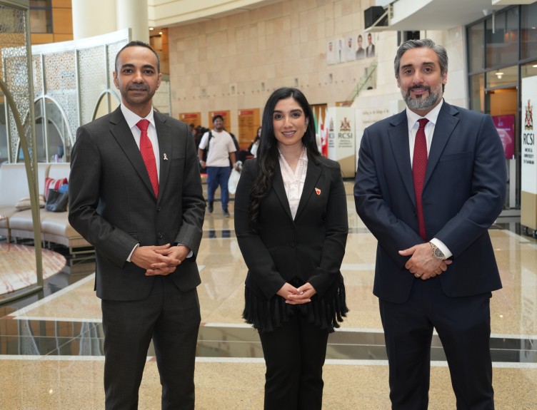
An important part of the project has been the collaboration behind it. The work brought together RCSI Medical University of Bahrain, Royal Medical Services - Mohammed bin Khalifa bin Salman Al-Khalifa Cardiac Center, Salmaniya Medical Complex and Family Medical Centre, with additional academic collaboration from Al Faisal University in Saudi Arabia. The project brought together expertise in cardiology, paediatrics, radiology and medical physics.
The study is a good example of what can be achieved through collaboration between institutions in Bahrain. It allowed us to look at liver stiffness alongside clinical, laboratory and haemodynamic features rather than in isolation.
This broader approach is likely to be important in FALD. Our findings suggest that 2D-SWE may be most useful as part of a wider assessment, rather than as a single measure of fibrosis. Looking at these measurements together may help clinicians tailor long-term follow-up, identify patients who need closer monitoring and decide when further imaging or specialist assessment is needed.
The next step is to understand how these measurements change over time and whether combining elastography with other clinical and imaging markers can improve long-term surveillance.
References
4. Abdulla M, Al-Hashimi H, Kalis N, Blackwell J. Discordant 2D shear wave elastography values in post-Fontan patients with ascites: a potential interpretive pitfall. Journal of Ultrasound. 2026