Researchers improve technique for growing lung cells

A team led by an RCSI researcher has improved how they generate new lung cells in a dish, which could lead to better treatments and cures for lung diseases in the future.
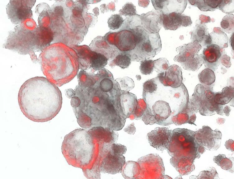
Using this technique, cells can be grown in a laboratory and stored for more than one year without losing their lung identity and can be used to model lung diseases.
The researchers were able to do this by using a combination of pluripotent stem cells (cells that can potentially produce any cell or tissue type) and machine learning (artificial intelligence that allows computers to learn automatically).
The new technique was developed in collaboration with Boston University and Carnegie Mellon University (CMU) in Pittsburgh. Their findings appear online in the journal Cell Stem Cell.
Induced pluripotent stem (iPS) cells are derived from the donated skin or blood cells of adults and, with the reactivation of four genes, are reprogrammed back to an embryonic stem cell-like state. iPS cells can be differentiated toward any cell type in the body and do not require the use of embryos.
Building on previous work from the Center for Regenerative Medicine (CReM) of Boston University and Boston Medical Center, the researchers reprogrammed blood from adults into iPS cells. They then treated these stem cells over a period of one month until they became cells which were very similar to adult lung cells.
According to the researchers, often when this type of experiment is performed the resulting cells are not a pure collection of the cell that they aimed to create (target cell) and they do not keep the characteristics of the target cell for prolonged periods of time.
“Therefore, we developed a combination of techniques that examines the gene expression of thousands of single cells combined with DNA barcoding of each individual cell and machine learning to build up a dynamic picture of what factors favor cells that go on to be lung cells in our system. Using this knowledge we were able to improve our methods for generating lung cells so that we can now create more relevant cells that keep their cell identity in a dish for more than one year,” explained Killian Hurley, MD, PhD, researcher at RCSI, who co-authored the study with Jun Ding, PhD, a post-doctoral fellow at CMU.
The researchers believe this study will improve their ability to model lung disease and treatments in the laboratory for diseases including idiopathic pulmonary fibrosis, chronic obstructive pulmonary disease (COPD), alpha-1 antitrypsin deficiency and neonatal respiratory distress or early-onset interstitial lung disease.
Millions of people around the world have severe lung diseases, often without good treatments or cure. Some of these diseases may even require lung transplantation which is a complex and high risk surgery with the need for donor organs always exceeding the supply.
“The machine learning methods we developed for this study can also be applied to studies of other tissues and organs,” said Ding. “We hope that our newly developed techniques for generating a pure, unlimited supply of cells using patients-derived stem cells can make possible new treatments or cures for diseases. These developments would prolong lives and improve the quality of those lives.”
“The key hurdle to understanding what goes wrong with an individual patient’s lung cells has been our inability to access those cells or to grow them in the laboratory. This approach allows us to now engineer from any individual patient those very finicky cells and to introduce bar codes into those cells that allow us to track and understand each cell and all their progeny over time in the laboratory dish. The result is an inexhaustible source of new lung cells that can be prepared from any patient of any age,” added co-corresponding author Darrell Kotton, MD, David C. Seldin Professor of Medicine and Director, CReM, who led the work together with Ziv Bar-Joseph, PhD, the FORE Systems Professor of Computer Science at CMU.
Funding for this study was provided by an Alpha-1 Foundation Postdoctoral Fellowship Award (KH), TL1TR001410 and F31HL134274 (AJ), F30HL142169 (YS), the I.M. Rosenzweig Junior Investigator Award from the Pulmonary Fibrosis Foundation (KDA), R01GM122096 and R01HL128172 (ZBJ and DNK), OT2OD026682 (ZBJ), and R01HL095993, R01HL122442, U01HL134745, and U01HL134766 (DNK).


