RCSI researchers develop first synthetic mitral valve model to replicate the heart's natural mechanics

Researchers at RCSI University of Medicine and Health Sciences have developed an artificial model of the mitral heart valve that faithfully mimics the valve’s complex mechanical behaviour in the human heart.
The study could help researchers better understand valve disease and develop new treatment approaches.
The mitral valve opens and closes around 100,000 times each day, making its mechanical properties critical to healthy heart function. When the valve does not work properly, blood can leak backwards through the heart in a condition called ‘mitral regurgitation’, which affects tens of millions of people worldwide. As global life expectancy increases, the number of people experiencing mitral valve conditions is expected to rise.
A new, low-cost mitral valve model developed by RCSI can replicate the behaviour of the ‘native’ mitral valve in the human heart, and so will enable researchers around the world to better understand how the mitral valve works and develop new approaches to restoring its function.
“Advancing our understanding of mitral valve function is dependent on developing synthetic alternatives that capture the valve’s complex mechanical behaviour, which is achieved in this study,” explained Dr Claire Conway, Lecturer in RCSI Department of Anatomy and Regenerative Medicine, and author of the new study published in Acta Biomaterialia.
Up to now, synthetic mitral valves have lacked anisotropy, or the property of having different directional mechanical properties, and such models could not withstand the levels of pressure and flow rates that are seen naturally in the human heart.
The new model is the first of its kind to incorporate the mechanical properties of real heart valve tissue while also operating under realistic heart pressures and flow conditions. And because many mitral valve problems are underpinned by alterations in mechanics, having a model that behaves like human tissue can provide a new window into how mitral malfunctions begin and progress.
“This model captures native anatomy and the fabrication is precise and repeatable,” said Dr Conway. “Physical and digital tests of the valve revealed it successfully functioned under physiological flow and physiological pressure, representing a significant advance in the field.”
Importantly, the new model also allows precise control over the tension and thickness of the leaflets that enable the mitral valve to open and close effectively.
“This model gives us precise control over key features of the mitral valve while still reproducing the way it functions in the heart," said Dr Sina Javadpour, first author of the study and Postdoctoral Fellow at Trinity College Dublin. "That makes it a powerful tool for studying valve disease and testing new repair strategies in a controlled laboratory environment."
The research was funded through a RCSI StAR Lectureship and the Research Ireland Frontiers for the Future Programme, and carried out by the RCSI Tissue Engineering Research Group.
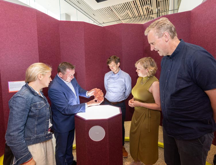
Members of the public can see the model valves as part of the Heart exhibition in Humanarium at RCSI.